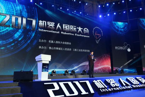
According to the latest data, 8.8 million people die of cancer every year, accounting for nearly one-sixth of the total annual deaths worldwide. Most of the deceased are in low- and middle-income countries. There are more than 14 million new cancer cases every year. It is expected that this number will increase to more than 21 million by 2030. Cancer has become a cloud in the world. On September 21st, at the “2017 Robot International Conference†jointly organized by the Sanhang Industrial Technology Research Institute in Foshan City, Professor of the City University of Hong Kong, doctoral tutor, head of the Department of Mechanical and Biomedical Engineering, IEEE Fellow Professor Sun Dong, an academician of HKIE, proposed cell surgery based on micro-operation and micro-robot technology, which has increased our expectation for future cancer treatment. 2017 Robot International Conference Professor Sun Dong said that the development of cancer disease has changed from a single cell to a relatively large tumor, from the first stage to the next stage, and finally to cancer cells. At present, our medical methods are mainly concentrated in the tumor stage, but at present we all hope to achieve early diagnosis and early treatment, mainly in single cells. If you want to achieve this, you need to be very precise. The treatment of the cells is done by surgery. We can change one cell, then inject the cells into the tumor cell cluster and change other cells. Our development of cell surgery over the past 20 years has been very long-term, and it has evolved from a very new nuclear cell concept to a recently successful clinical trial, which is a successful case of our single-cell surgery in 2016. Another very successful case is the treatment of leukemia by changing genes. We treat leukemia by changing the genes of cells in patients. Now we mainly face the problems of low precision, low efficiency, insufficient continuity, low success rate, insufficient proficiency, etc., and now we hope that it can achieve more comprehensive detection, higher success rate, and more. Good image feedback and artificial intelligence, etc. For these operations, we want robots to help us achieve these goals. What is a cell surgery robot? Traditional medical robots have risen from the organ level to the cellular level, even reaching subcellular levels. We now have three different operating arms for single-cell operation. The first is VIVO, called laser projection single-cell surgery, which treats lasers in a blood cell or red blood cell. The second is that the operation of the microneedle can perform single cell surgery. The third is cell modification technology. Hong Kong now has a team to conduct a new research, automated optimization of cell operators, the ability to deliver cells to subtle organs through the manipulator, and then integrate the two to form a preliminary service robot. Through sophisticated delivery, we can improve the technology of robots in this area. The global robot market is growing rapidly, with an annual growth of more than 20%. The world will grow by 32 billion next year. One is the medical robot market, the other is a cellular robot, and there will even be higher demand for cell surgery. For cell therapy in automated mechanical operators, Sun Dong said that the therapy is based primarily on the foundations of several research techniques, such as optical operators, micro-organization operators, 3D operations, video control, video distribution, and microchip technology. The system allows for the transplantation of single cells in vitro and in vivo. We can do some live needle testing and microinjection within and outside our vision plan. By tracking the particles, in a very detailed way, these particles have a tiny volume of 10 microns to 10 mm. We believe that optical speculation can make the robot perform proficient operations and become a skilled operating tool that can perform on cells. Contrast, high flexibility and high mobility. What I said earlier is the mechanical and laser-based measures system. What can we do after we fuse the cells? We need to transform the cells into new types. We need automated operators for fine delivery. Fine medical treatment can be applied to tissue repair, tissue regeneration, etc. This is the problem faced by our engineers. There are two challenges here. First, we need to develop a device that allows micro-materials to enter the body. This system must provide a lot of work space. We need particle control in 3D, and now only a few research groups around the world can produce such equipment. The second challenge is to design a porcelain robot that moves the robot inside the body. We are now developing the second edition, which is now available in 100 micron, 200 micron to 300 micron workspaces, which is large enough to enable the task. In the new version, we can detect these particles. When you design and operate the robot, our robot has a porous structure, in this way we can have enough channels for nutrient delivery. Its spiral structure helps our robots attach and transport, and these cells can successfully grow on the robot. As for how to apply 3D printing technology? Professor Sun Dong said that the design of 3D printing technology is carried out in the laboratory. With the laser scanning technology, different methods are used. Our small robot is only 80 microns in size. Now our VIVO transportation experiment can be successfully performed on one cell. Transporting our robots, there are many visual and dynamic controls that control its movement and trajectory with great precision. We can successfully control the movement of this embryonic cell and move to where we want it to go, very precise. We have done this test many times to ensure that the accuracy is sufficient and all precision errors are zero. The next thing we want to consider is whether the robot can be separated from the cell. This is very important because we can move the cells to a place. How can we let the cells leave the tissue and leave the organ through the operation of the robot? We now assume that there is a structure that helps the cells leave the tissue, we leave it at the same time as we test, and now we are experimenting with mice. After a week, we can see that the cells really start to grow, start to replicate, and finally become cancer cells. Then we find that the cells are released from the robot. We use this mouse to experiment to study cancer and stem cells in white mice. The development of the body. Cancer is terrible, terrible ailments, and with the development of future technologies, cancer may become a common condition, and it may be completely cured by simple surgery. Hydrocolloid Applicator,Hydrocolloid Dressing,Hydrocolloid Band Aid,Hydrocolloid Gel Patches Henan Maidingkang Medical Technology Co.,Ltd , https://www.mdkmedical.com