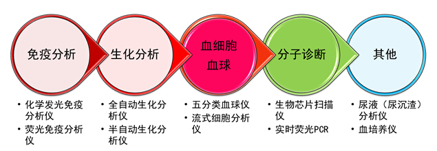
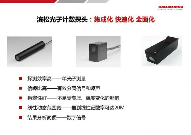
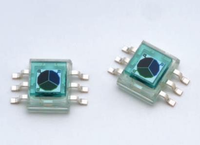
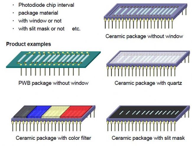
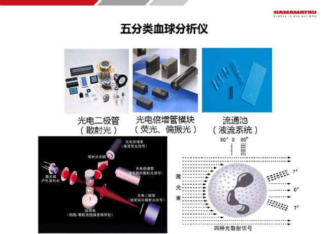
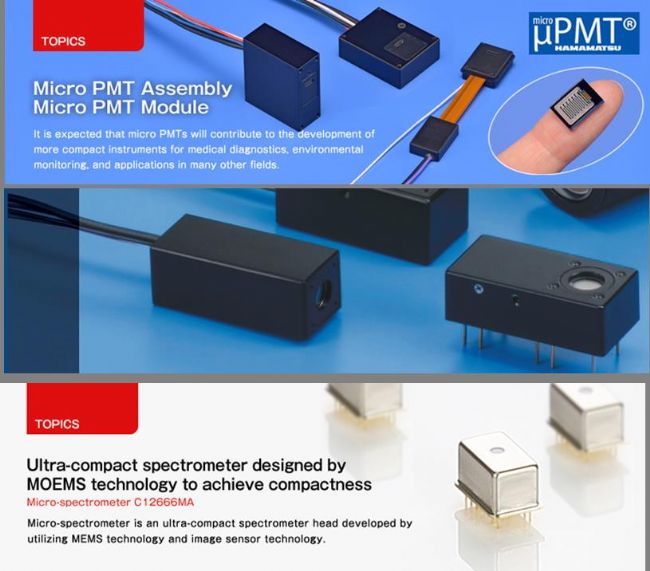
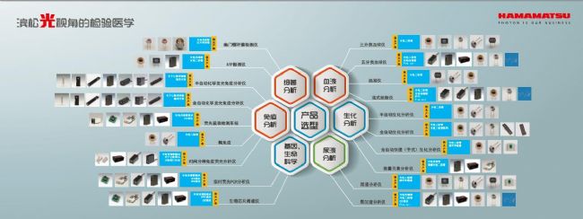
In Vitro Diagnosis (IVD) is a diagnosis in which blood, body fluid, and tissue samples are taken out of the human body and then detected. IVD plays an increasingly important role in modern society, and more than 80% of clinically diagnosed diseases rely on it. It plays an extremely important role in the whole process of disease prevention, diagnosis, monitoring and guiding treatment. It is an indispensable tool for modern disease and health management, and is also known as the doctor's "eye". [1] What makes these big "eyes" truly capable of discovery and diagnosis is the "light" in their hearts, which is the "optical method" used in in vitro diagnostics. In the traditional laboratory, we can analyze the type and quantity of the material by the characteristics of emission and absorption of the spectrum; use the scattering characteristics of the particles to analyze the size and amount of the particles. Nowadays, the traditional methods of analytical instruments are combined with hematology, immunology, biochemistry, and labeling techniques to form the current method of clinical testing. The optical method is a safe, reliable, non-contact detection method. Testing fluorescence, analyzing spectra, reading colors, etc. are typical applications of optical methods in laboratory medicine. Laboratory medical equipment typically using optical methods Just like in the traditional analytical instrument industry, photodetectors and light sources also play an important role in in vitro diagnostic instruments. Don't look at our detectors may be just a small piece in a device, but it is a methodological At the core, the performance of the detector plays a key role in the realization of the function of the device and the performance. Laboratory medical equipment generally has high requirements for sensor detection limits, signal-to-noise ratio, dynamic range, and anti-jamming performance. So, what are the types of "optical methods" in in vitro diagnostics? How do photodetectors, light sources, etc. correspond to related applications? Next, let us take a closer look at the in vitro diagnostic technology under the "light perspective" and uncover the answers to these questions! First, the luminescence method At present, the main types of luminescence in in vitro diagnosis are as follows 1. Chemiluminescence refers to the phenomenon of emission of light generated by a chemical reaction process. Some substances (light-emitting agents) absorb the chemical energy generated during the reaction in the chemical reaction, causing the electrons in the reaction product molecules or the intermediate molecules of the reaction to transition to the excited state, and when the electrons return from the excited state to the ground state. At the time, energy is released in the form of emitted photons, a phenomenon known as chemiluminescence. Among them, there are enzymatic reactions, electrochemiluminescence, etc., such as chemiluminescence immunoassay analyzers and electrochemiluminescence immunoassays. 2. Bioluminescence refers to the phenomenon of luminescence occurring in living organisms, such as the luminescence of fireflies. The reaction substrate is firefly luciferin. Under the catalysis of luciferase, ATP energy is used to generate excited oxidized fluorescein. It releases excess energy in the form of photons when it returns to the ground state. We can use this method to detect some viruses and bacteria. The glow of fireflies belongs to bioluminescence 3. Photoluminescence, which is often referred to as fluorescence, means that the illuminant (fluorescein) is excited by a short-wavelength incident light, and the electron absorbing energy transitions to an excited state, and when it returns to the ground state, it emits A longer wavelength of visible light (fluorescence). This method is used for real-time fluorescent PCR, flow cytometry, time-resolved immunofluorescence, and biochip scanners that are often heard. Using immunological and genetic methods, we add specific markers to the blood, body fluids, tissues, etc. to be tested, and these markers emit specific reactions in the presence of chemical reactions, enzymatic reactions, currents, or illumination. The light, we can get the number of markers by detecting the intensity of the light, to determine how much material is contained in the sample. This method is currently used in chemiluminescence immunoassays, electrochemiluminescence analyzers, flow cytometry Analyzer, fluorescent PCR and other instruments. Detector and light source: Photomultiplier tube modules and photon counting probes are playing an irreplaceable role as very weak light detection devices, and flashing xenon lamps are increasingly favored by instrument manufacturers as high-power ultraviolet light sources. Second, colorimetry Colorimetry, the same is true, after the reaction of the label with a specific reagent, there will be a color reaction, like the PH test paper we are familiar with, will show different colors under different pH. The same is true for in vitro diagnostics. For example, the gold standard instrument and the urine analyzer are typical color developments. Different substances and different substance contents will make the test paper or reagent display different colors. For the traditional method, we can judge the color through our eyes. However, under different people and different light conditions, the human eye will have a deviation in color resolution, which is subjective and consumes a lot of manpower. Now People use optical methods to make different reflections of light with different colors. Detector and light source: We can measure the amount of light reflection by color sensor or photodiode, and then we can get the color information and effectively improve the accuracy of the inspection. Color sensor Third, the spectral absorption method Spectral absorption method is a typical method for analyzing the type and amount of substances. It is widely used in laboratory equipment. It has atomic spectrum, molecular spectrum, absorption spectrum, emission spectrum, etc. The biochemical analyzer used in in vitro diagnostics uses this classic. The analysis method analyzes the contents of different substances such as serum, urine, and cerebrospinal fluid. Biochemical analyzers can be viewed as spectrophotometers for special applications. Detector and light source: Since it is a spectrophotometer, many optoelectronic components can be used, such as light sources, photomultiplier tubes, CCD chips, micro spectrometers, and so on. However, biochemical analyzers are used to analyze specific substances. Generally, there are only 10-16 channels. Photodetectors or photodiode arrays can be used in this detector. It is suitable for single-point photodiodes of slower test equipment. of. For high-end, high-speed test equipment, you need to customize the photodiode array according to the required spectral position. Custom Photodiode Array for Biochemical Analyzers Fourth, scattering method Scattering is commonly used to determine the size and size of particulate matter. The same method can be used to analyze cell size and structural information. For example, the five-class hemocytometer and urine sediment analyzer use this traditional method. Of course, in equipment such as five-class blood cells, urine sediments, etc., in order to obtain more characteristics about cells and other formed components, the method of the luminescence method we mentioned is also used. Detector and light source: In this method, photodiodes, photomultiplier modules, and flow cells will play a very important role. In recent years, due to the many needs of diagnosis, treatment, monitoring and medical research in clinical medicine, the development of medical testing methods has been very rapid. Any new theory, new technology in hematology, immunology, biochemistry, microbiology, optics, computer science, any which can be used to diagnose diseases, will sooner or later develop into a test methodology and be applied to clinical or laboratory. Optical applications in other in vitro diagnostics The new development has brought new demands. At present, in vitro diagnostic equipment is developing in two directions, which is large-scale, rapid, and comprehensive in function, corresponding to portability, simplification, and specialization. Hamamatsu's miniaturized, integrated, low-power photodetector solution No matter which direction of development, or which in vitro diagnostic application, as a supplier of core photodetectors, in addition to the need to ensure the stability of the most basic supply and product performance, and to provide reasonable customers In addition to product solutions and differentiated customized services, it is also important to share a broader vision of industry development, and it must be done. Hamamatsu's "Light Perspective" Laboratory Medicine The above products are all optoelectronic components of Hamamatsu. Note: [1] quoted from "in vitro diagnostics on the tuyere" Water Hose Cart,Water Hose Reel Cart,Garden Water Hose Reel Cart,Industrial Water Hose Reel Cart NINGBO QIKAI ENVIRONMENTAL TECHNOLOGY CO.,LTD , https://www.hosereelqikai.com