American research team successfully realized the imaging of whole brain nerve activity in Drosophila larvae
Release date: 2015-08-17
Every second of your brain and nervous system has millions of nerve signals flowing, allowing you to do everything in your life. Now that this process can be captured for the first time, scientists have used advanced microscopic imaging techniques to capture the neural signal activity of Drosophila larvae for the first time.

The video shows the process of neuronal firing of the nervous system as the larva crawls and climbs, and the results are amazing. The system captures five times a second and captures the discharge of a single neuron in an hour. It is impossible to capture the brain activity of complex organisms such as fruit flies in this way.
Scientists at the Janelia Research Camp at the Howard Hughes Medical School in Virginia, USA, used light microscopy to study the central nervous system of millimeter-length Drosophila larvae.
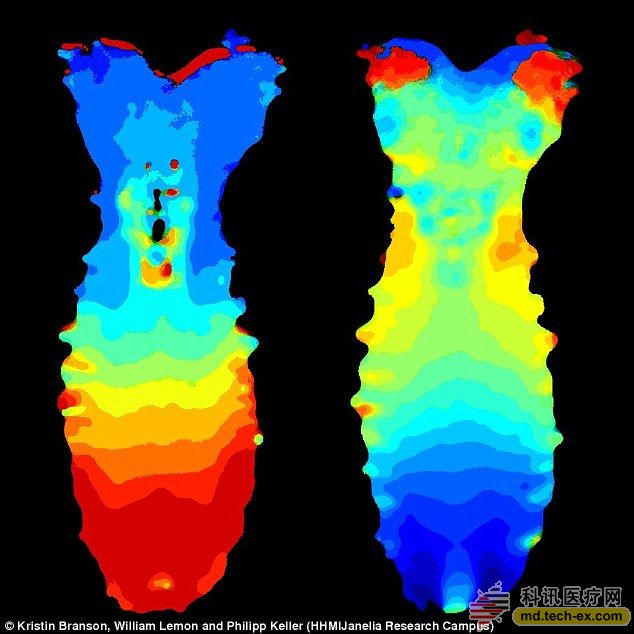
The Drosophila sample is illuminated from both sides of the laser and the camera records the image information. The researchers modified the neurons by genetic techniques to allow them to glow when they were discharged. They also stripped the heatstroke nervous system from the body of the larva for easy viewing.
The system successfully recorded that neural activity showed how the Drosophila larva nervous system signals its body to move.
The team made great progress from observing the transparent zebrafish to the current fruit fly in 2013, because the fruit fly is opaque and the central nervous system is more complicated.
Researchers believe that brain-wide imaging technology allows scientists to look at how the brain works from a better perspective, and their next goal is to image the neural activity of mouse embryos.
Original link: http://
Source: Kexun Medical Network
Colored adhesive bandage
Wenzhou Celecare Medical Instruments Co.,Ltd , https://www.celecaremed.com