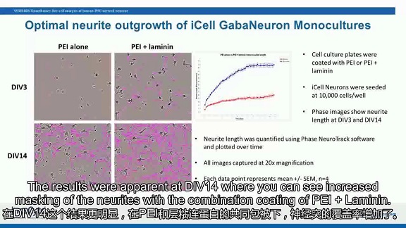
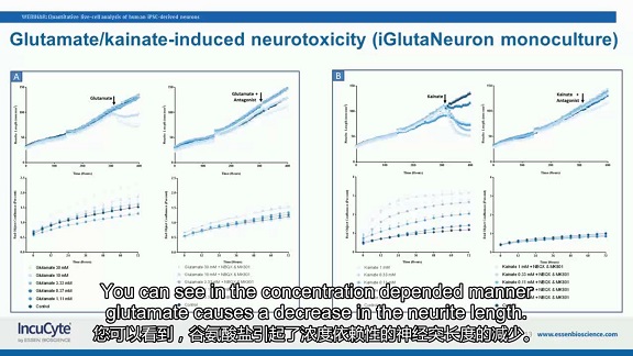
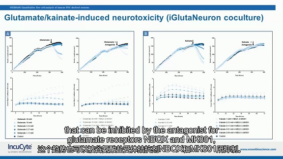
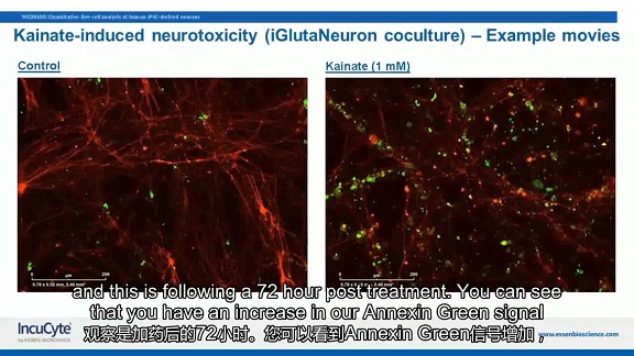
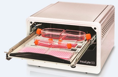
I. Introduction One of the main limitations in studying the effects of human disease on the nervous system is the ability to culture, monitor, and analyze nerve cells to accurately represent the phenotype of these diseases. Research on human induced pluripotent stem cell (hiPSC)-derived neural cells provides a new method for the modeling of neurological diseases. How to monitor the morphology and cellular health of neurons in long-term culture is the key to describing and evaluating these new model systems. Traditional experimental/imaging techniques that rely on endpoint methods require immunochemical staining and can only provide limited real-time dynamic information. In this webinar, we present a study of IncuCyte S3 for long-term dynamic quantitative analysis of human iPSC-derived neurons. Second, the lecture introduction This lecture describes how to optimize the culture conditions of human iPSC-derived neurons using IncuCyte's Live Cell Dynamic Imaging and Analysis System ; how to perform real-time live cell analysis using phase contrast imaging (individual culture) or fluorescence imaging (co-culture); NeuroTrack software images and quantifies the dynamic changes in neurites; how to use other live cell phenotypic experiments (such as "apoptosis") to monitor neuronal health and validate its mechanisms of activity. Figure 1. Lecture title Third, the experimental method Our experiments are divided into single culture and co-culture. 3.1 Using single culture to study culture conditions and adhesion of iPSC-derived neural cells to the culture plate We inoculated nerve cells (iCell GabaNeurons) on PDL, PLO or PEI, and the plates were coated twice with or without Matrigel or Laminin. The IncuCyte S3 was then placed and analyzed by phase difference NeuroTrack software for quantification of neurite length growth over time. 3.2 Study the effects of neurotoxic compounds by co-culture Using glutamate nerve cells (iGlutaNeuron) and primary rat astrocyte co-culture system, nerve cells were plated on day 1 (DIV0), IncuCyte NeuroLight Red reagent was added for red lentiviral transfection and placed in IncuCyte In S3; these agents were removed on day 2 (DIV1), astrocytes were plated, placed again in IncuCyte S3, and neurite outgrowth was tracked using the fluorescent NeuroTrack algorithm. A neurotoxic compound, or a compound and its receptor antagonist, was added on day 15 (DIV14), and multiplex reaction was performed by adding Annexin Green reagent for monitoring apoptosis, followed by monitoring for 72 hr. Fourth, the experimental results 4.1 Studying culture conditions with single culture and adhesion of iPSC-derived neural cells to the culture plate Cells implanted on PDL or PLO form large neuronal globules regardless of the presence of additional matrigel or laminin coating. However, in the presence of Matrigel or Laminin coating, neurites grow more than PDL or PLO alone. For cells implanted on PEI, under the co-coating of PEI and laminin, neurite outgrowth is more robust and coverage is increased (Figure 2). Figure 2. The image on the left shows that at the time of DIV3, under the common coating of PEI and laminin (PEI+laminin), neurite growth is more robust; at DIV14, in PEI and laminin (PEI+laminin) Under the common contraction, the coverage of neurites has also increased. In the graph on the right, the blue line indicates PEI+ laminin, the red line indicates individual PEI, and the blue line indicates that the neurite length increases faster with time than the red line. 4.2 Study the effects of neurotoxic compounds by single culture and co-culture We will study excitotoxicity and study how the neurotoxic compounds Glutamate and Kainate can affect neurite outgrowth. For the glutamate neuron (iGlutaNeuron) culture system alone, we labeled the living neurons with a nuclear marker (IncuCyte NeuroLight Red) and monitored the neurite outgrowth with IncuCyte S3 for 14 days. The length of the protrusion increases significantly with time. On the 15th day, we added glutamate at different concentrations. Glutamate caused a decrease in concentration-dependent neurite length , which was inhibited by the glutamate receptor antagonists NBQX and MK801 (two images in Figure 3A). We also added the Annexin Green reagent to monitor apoptosis for multiplex reaction, and the apoptotic cells will be stained with green fluorescence. After glutamate treatment, an increase in glutamate concentration can cause an increase in the signal of Annexin Green reagent, indicating an increase in apoptosis, which is also inhibited by the glutamate receptor antagonists NBQX and MK801 (Fig. 3A next two pictures). Addition of kainate can also give similar results to the addition of glutamate: the addition of kainate can cause a concentration-dependent decrease in neurite length and an increase in the signal of Annexin Green reagent. This trend can also be Inhibited by the glutamate receptor antagonists NBQX and MK801 (Fig. 3B). Figure 3. iGlutaNeuron nerve cells cultured separately. The left part A indicates the neurotoxic effect of glutamate: The upper left image shows the addition of glutamate at the time of DIV14, and the neurite length is reduced. The higher the glutamate concentration, the more the neurite length is reduced. In the upper right picture, the simultaneous addition of glutamate and glutamate receptor antagonist (Antagonist) at the time of DIV14, the tendency of neurite length reduction was alleviated. The lower left panel shows that glutamate can cause an increase in the signal of the Annexin Green reagent, and the higher the glutamate concentration, the more the signal increases. The lower right panel shows that an increase in the signal of the Annexin Green reagent can also be inhibited by a glutamate receptor antagonist. The B part on the right side shows the neurotoxic effect of kainate: the experimental results are similar to the A-part glutamate. In the co-culture system of glutamate nerve cells and primary rat astrocytes, we also studied how the neurotoxic compounds glutamate and kainate can affect the growth of neurites and get A similar conclusion (Figure 4). Figure 4. iGlutaNeuron nerve cells and astrocytes co-culture. Explain the same as Figure 3. This procedure is also supported by images, such as an increase in the signal (green fluorescence) of the Annexin Green reagent and a decrease in red fluorescence showing the length of the neurite (72) after 72 hr after the addition of kainate. Figure 5. 72 hr after the addition of kainate, an increase in the signal (green fluorescence) of Annexin Green reagent and a decrease in red fluorescence showing neurite length compared to the control (left panel) (right panel) . In summary, the lecture demonstrates how to use IncuCyte S3 for dynamic cell analysis, with video to provide truly valuable visual information and validate quantitative data from experiments, greatly advancing insights into biological research. V. Automated live cell dynamic analysis At present, most of the cell detection methods still belong to the traditional end point method - only the final result is given. This method can not only dynamically monitor and analyze the whole process of cell experiments, but also obtain biased results. In order to be able to observe live cells throughout the process, Essen Corporation of the United States has developed the IncuCyte "Viable Cell Dynamic Imaging and Analysis System" built into the incubator, inside the incubator, and simultaneously for 6 cell culture dishes/bottles/microplates (6- 384-well) for long-term dynamic imaging, recording real-time changes in cell morphology details, and transforming into a variety of cell-related time-quantitative curves through powerful image analysis software. Figure 6. IncuCyte S3: Live Cell Dynamic Imaging and Analysis System IncuCyte is a cell-level scientific and drug screening solution that has been used for immunosuppression, nerve growth and regression, cancer cell metastasis, cell migration, stem cell differentiation, apoptosis, cell necrosis, cell proliferation, wound repair, etc. Scale screening and research on long-term life science issues. Sixth, online lecture video Video duration is 22min, please watch under WiFi conditions: Https://v.qq.com/x/page/y0513k5eicg.html 7. About "Diaoao Biotechnology" “ Diao Biotech †(Shanghai, Hong Kong, Taiwan) was established in 1994. For more than 20 years, it has been adhering to the world-class instrument manufacturer. The experts with rich R&D experience are technical support and the professional maintenance team serves. Backed by a focus on providing state-of-the-art hardware and software equipment and technical services for life sciences, pharmaceutical R&D and disease treatment. The exclusive distributors of " Diao Biotech " are: IncuCyte, Beacon, Gyrolab, Chipcytometry, iQue Screener, Avatar, PlexBio, Certus, HighRes, etc. Telephone (transfer to the marketing department) Email: Website: Scan the QR code to add the WeChat public account " tekontech ": Inflation Devices,Cardiology angiography,PTCA balloon inflation devices,coronary angiography Anesthesia Medical Co., Ltd. , https://www.jssinoanesthesia.com